image


Clemmer Group

Protein Dynamics in an Ion Trap


image
Figure 1: Cytochrome c ions held within an ion trap for varying lengths of time from 0–150 ms. Data a plotted on a nested drift time(flight time) scale where drift times correspond to ion mobility, and flight times correspond to m/z ratio as measured by a time-of-flight mass spectrometer. Here, we can see the 7+ charge state of cytochrome c at the time (longer flight times) and the 15+ charge state at the bottom (shorter flight times).

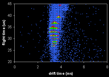
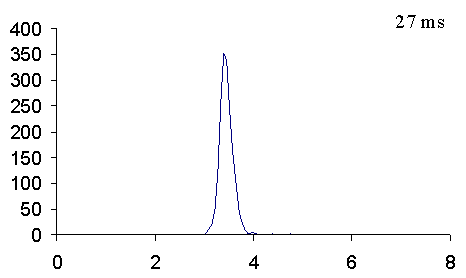
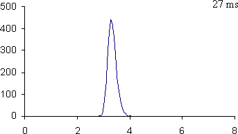
As ions are stored in an ion trap for longer periods of time, the observed distribution of conformations for each charge state shifts from relatively compact states (low drift time) to much more open conformations (high drift time).


image
Figure 2: Cytochrome c 8+, shown as a function of signal intensity on a drift time (in ms) scale.


image
Figure 3: Cytochrome c 9+, shown as a function of signal intensity on a drift time (in ms) scale.


Badman, E. R. and Clemmer, D. E., unpublished results.